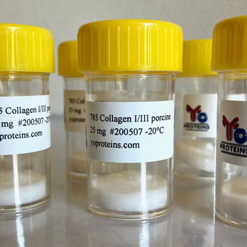
889 Collagen type III (human) 0.1 mg
0 EUR
249 EUR
Collagen type III protein purified from human placenta
Source
Human placenta. Negative for HBsAg, HCV, HIV1 and HIV2
Purity
Human collagen type III at least 90%, human collagen type I below 10%, other human collagen types and non-collage proteins below 1%
Format
Lyophilized
Purification
Partial pepsin digestion in acidic conditions, differential salt precipitation and DEAE chromatography
Applications
Collagen type III standard (e.g. for cellular assays, ELISA, WB and dot blotting) (3) (4) (9) (10) (11)
Antigen for antibody production. Coating material for cell culture studies
Detection of IgGs against disc ECM proteins by immunoblotting (2)
Collagen was used to polarize THP1 monocytes differentiation (5)
Cell attachment in conjugated ECM-PA microarrays (6) (7)
Use of collagen for fabrication of cell microarrays that were used for studying drug response on lung adenocarcinoma cells (8)
Reconstitution
Use 0.5 M acetic acid, pH 2.5
Storage
Shipping at ambient temperature. Storage 2 years at -20�C or lower
Reference
1. Wegener H, Leineweber S, Seeger K. (2012) The vWFA2 domain of type VII collagen is responsible for collagen binding. Biochem Biophys Res Commun. Dec 10 PMID: 23237810
2. Capossela S, Schläfli P, Bertolo A, Janner T, Stadler BM, Pätzel T, Baur M, Stoyanov JV. Degenerated human intervertebral discs contain autoantibodies against extracellular matrix proteins. Eur Cell Mater. 2014 Apr 4;27:251-63 PMID: 24706108
3. Philips N, Chalensouk-Khaosaat J and Gonzalez S (2015) Stimulation of the Fibrillar Collagen and Heat Shock Proteins by Nicotinamide or Its Derivatives in Non-Irradiated or UVA Radiated Fibroblasts, and Direct Anti-Oxidant Activity of Nicotinamide Derivatives. Cosmetics 2015, 2(2), 146-161; doi:10.3390/cosmetics2020146
4. Kuzan A, Chwilkowska A, Maksymowicz K, Szydelko-Bronowicka A, Stach K, Pezowicz C, Gamian A (2018) Advanced glycation end products as a source of artifacts in immunoenzymatic methods. Glycoconjugate Journal 35(1) DOI: 10.1007/s10719-017-9805-4 PMID: 29305778
5. Pal S, V. Badireenath Konkimalla (2016) Data on sulforaphane treatment mediated suppression of autoreactive, inflammatory M1 macrophages. Data in Brief. https://doi.org/10.1016/j.dib.2016.03.105 PMID: 27222853
6. Kaylan KB, Ermilova V, Yada RC and Underhill GH (2016) Combinatorial microenvironmental regulation of liver progenitor differentiation by Notch ligands, TGF, and extracellular matrix. Scientific Reports volume 6, Article number: 23490 PMID: 27025873
7. Kourouklis AP, Kaylan KB, Underhill GH (2016) Substrate stiffness and matrix composition coordinately control the differentiation of liver progenitor cells. Biomaterials 99:82-94 PMID: 27235994
8. Kaylan KB, Gentile SD, Milling LE, Bhinge KN, Kosari F and Underhill GH (2016) Mapping lung tumor cell drug responses as a function of matrix context and genotype using cell microarrays. Integrative Biology. Issue 12 PMID: 27796394
9. Meschiari CA, Jung M, Iyer RP, Yabluchanskiy A, Toba H, Garrett MR, Lindsey ML (2018) Macrophage overexpression of matrix metalloproteinase-9 in aged mice improves diastolic physiology and cardiac wound healing after myocardial infarction. American Journal of Physiology https://doi.org/10.1152/ajpheart.00453.2017 PMID: 29030341
10. Sarah D. Ackerman, Rong Luo, Yannick Poitelon, Amit Mogha, Breanne L. Harty, Mitchell D'Rozario, Nicholas E. Sanchez, Asvin K.K. Lakkaraju, Paul Gamble, Jun Li, Jun Qu, Matthew R. MacEwan, Wilson Zachary Ray, Adriano Aguzzi, M. Laura Feltri, Xianhua Piao, Kelly R. Monk (2018) GPR56/ADGRG1 regulates development and maintenance of peripheral myelin. Journal of Experimental Medicine Mar 2018, 215 (3) 941-961; DOI: 10.1084/jem.20161714 PMID: 29367382
11. Smeulders N, Woolf AS, Wilcox DT (2003) Extracellular Matrix Protein Expression During Mouse Detrusor Development. J. Pediatric Surgery. Vol. 38, No 1 PMID: 12592609


